Bassano B., Durio P., Gallo Orsi U., Macchi E eds., 1992. Proceedings of the 1st Int. Symp. on Alpine Marmot and genera Marmota. Torino.
Edition électronique, Ramousse R., International Marmot Network, Lyon 2002

Anomalous tooth growth in an alpine marmot (Marmota marmota)

Barasa A. °, Galle Orsi U. * & Durio P. *
° Dipartimento di Morfofisiologia, Università di Torino. 1-10126, Torino.
* Dipartimento di Produzioni Animali, Epidemiologia ed Ecologia, Università di Turino. 1-10126, Torino


The skull, belonging to an adult Marmot (Marmota marmota), has the upper incisor which has over-grown because its masticatory surface has not been worn. Possibly this was caused by the corresponding lower incisor irregularly directed or congenitally absent. As a consequence the left tooth, coming through its socket, took the shape of a 340° arch. The growing direction is correct since normal upper incisors tend to converge toward the sagittal plane with an angle of about 20°.
Keeping growing the tooth reached the upper right jaw, perforated the mucosa, the periosteum and invaded the maxillary sinus in which a second and artificial, 9 mm-deep, tooth socket was created.
The infra-orbital duct, in which the hononymuos artery and nerve pass (brances of the intemal maxillary artery and of the maxillary nerve respectively) is crossed in an acute angle by the portion of the tooth pushed in the maxillary bone. Anyway the infra-orbital duct's width is not reduced when compared with the one on the opposite side.
The wall of the natural socket of this tooth is thicker and its internal surface is more irregular and rough when compared with the relatively smooth right upper incisor's socket.

Observation with optical microscope

The structure of thie bony tissue of the left tooth socket is more heterogeneous than the one of the opposite side (control). The bigger ammount of primary bony tissue and the lower degree of structural renewal observed indicate that the bony deposition was still going on.
Micro-hardness test using Vickers's procedure (4-faced pyramidal penetrator with 136° vertex angle) confirmed the high structural heterogenity, expressed by a wide dispersion of the obtained values.
Unfortunately the skull is missing the right incisor (which is was certainly present in vivo) and the whole mandible. Thus is not possible to understand why the tooth grow so much probably making mastication very difficult and eventually leading to starvation.
The tooth have been worn on the latero-lingual face and so we can assume that at least one lower incisor was present during the life of the animal.


Crescita dentaria anomala in una marmota alpina

Barasa A. °, Galle Orsi U. * & Durio P. *
° Dipartimento di Morfofisiologia, Università di Torino. 1-10126, Torino.
* Dipartimento di Produzioni Animali, Epidemiologia ed Ecologia, Università di Turino. 1-10126, Torino


Il cranio, appartenente ad un adulto di Marmotta (Marmota marmota), presenta l'incisivo superiore sinistro cresciuto più della norma in quanto la sua superficie masticatoria non é stata consumata. Probabilmente questo é dovuto all'incisivo inferiore corrispondente non diretto regolarmente o assente per cause congenite. Di conseguenza il dente sinistro emergendo dal silo alveolo ha assunto la forma di un arco di 340°. La direzione di crescita é corretta dal momento che gli incisivi superiori tendono normalmente a convergere verso il piano sagittale con un angolo di circa 20°. Mantenendo questa direzione di crescita il dente ha raggiunto la mascella destra, perforando la mucosa, il periostio e ha infine invaso il seno mascellare destro nel quale si é venuto a formare un falso alveolo della profondità di 9 mm. Il dotto infraorbitale, all'interno del quale decorrono l'arteria e il nervo omonimi (rispettivamente rami dell'arteria mascellare interna e del nervo mascellare), é attraversato, con un angolo acuto, dalla porzione del dente premente contro l'osso mascellare. Comunque la larghezza del dotto non é ridotta se paragonata a quella del versante opposto. La parete dell'alveolo naturale di questo dente é più spessa e la sua superficie interna é più irregolare e ruvida rispetto a quella dell'alveolo superiore destro.

Osservazioni al microscopio ottico

La struttura del tessuto osseo dell'alveolo dentale sinistro é pili eterogenea di quella controlaterale.La maggior crescita del tessuto osseo primario éd il minor grado di rinnovamento strutturale osservato indica che la deposizione ossea era ancora inatto al momento della morte dell'animale. Il test di microdurezza secondo la procedura di Wickers (penetratore piramidale a 4 facce con vertice a 136°) ha confermato l'elevata eterogenicità strutturale, espressa dall'ampia dispersione dei valori ottenuti. Sfortunatamente il cranio manca dell'incisivo superiore destro pur presente sucuramente in vita e dell'intera mandibola. Non é quindi possibile stabilire coma mai il dente sia cresciuto tanto fino a rendere la masticazione molto difficoltosa e portando probabilmente l'animale all'inedia. Il dente presenta segni di consunzione sulla faccia latero-linguale e possiamo quindi accertare la presenza nell'animale in vita di almeno un incisivo inferiore.

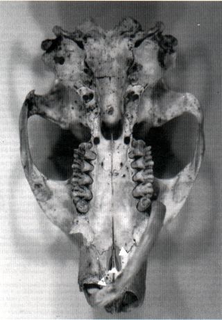
Plate 1: Ventral view

Â